You can access lymphoma patient models and PET simulation software, here: (GitHub).
For any questions, recommendations, or feedback, please contact: rfedrigo@student.ubc.ca
The upgraded XCAT phantom is available upon request from Dr. Paul Segars: paul.segars@duke.edu and additional information can be accessed at: https://olv.duke.edu/industry-investors/available-technologies/xcat/.
Summary:
- Novel lymphatic system was defined for the 4D-extended cardiac torso (XCAT) phantom
- A pipeline was developed to simulate and reconstruct PET images from the XCAT phantom, applying header information that is compatible with clinical radiology software
- As example application, simulated lymphoma patients were modelled using XCAT phantom and can be freely accessed
Reference:
Please use the following references if you publish results with help from this software tool:
R. Fedrigo, et al., "Development of scalable lymphatic system in the 4D XCAT phantom: Application to quantitative evaluation of lymphoma PET segmentations", Med. Phys., 2022
The Matlab component of this framework is adapted from the PET simulation and image reconstruction tool, as described by:
S. Ashrafinia, et al., “Generalized PSF modeling for optimized quantitative-task performance”, Phys. Med. Biol., vol. 62, pp. 5149-5179, 2017.
Technical Description:
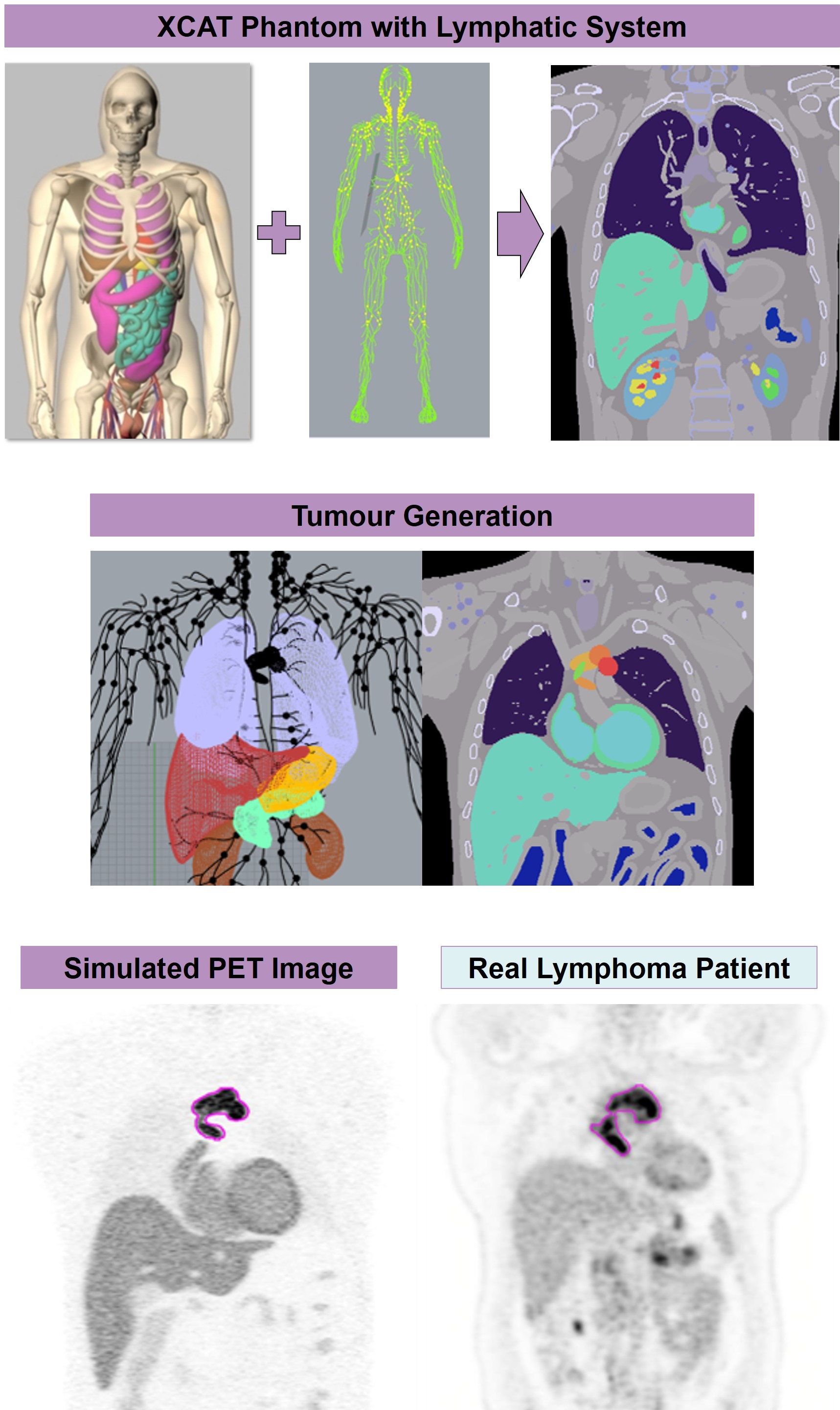
The novel lymphatic system for the 4D-extended cardiac torso (XCAT) phantom enhances the ability to model diseases, such as lymphoma. The lymphatic system (nodes, vessels) was defined using non-uniform rational basis spline (NURBS) surfaces. Multichannel large deformation diffeomorphic metric mapping (MC-LDDMM) method was used to propagate from the template phantom to different XCAT anatomies. The XCAT general parameter script was used to generate files that define the ground truth radioactivity and attenuation for a simulated patient.
Example files are provided, which can be used to model patients with non-Hodgkin’s lymphoma. Bulky tumours were created by altering lymph node morphology and function (nodes were expanded, stretched, converged, and had increased tracer uptake). This methodology is highly flexible and may be used in virtual clinical trials to simulate nodes with different pathology.
A framework was developed in Matlab and Python to simulate and reconstruct PET images using patients modelled with the XCAT phantom. Output files are converted to dicom and necessary header information is applied, such that the images can be viewed using clinical radiology software.