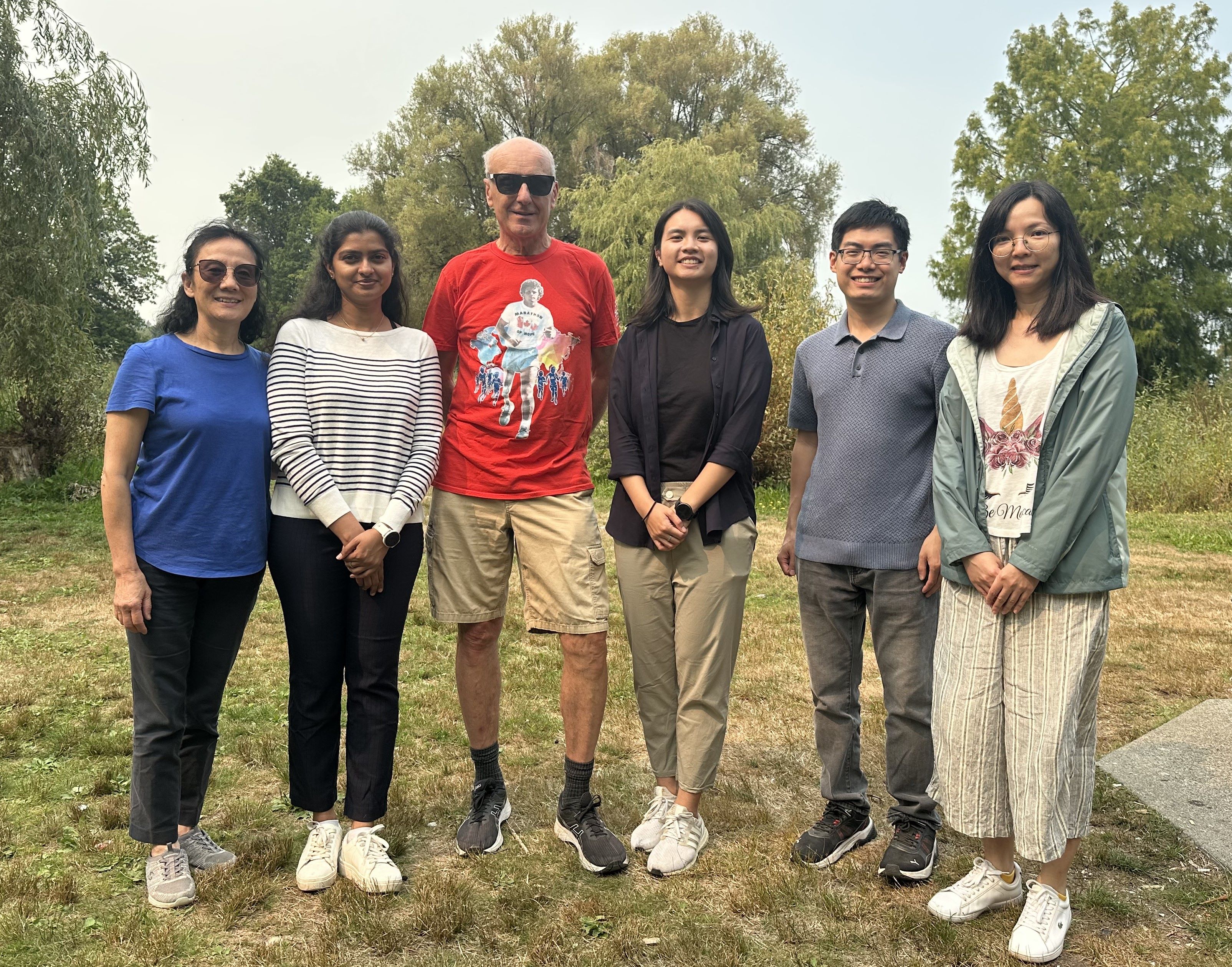
Lansdorp lab September 2025. From left to right: Yanni Wang, Rashika Ranasinghe, Peter Lansdorp, Tiffany Leung, Daniel Chan and Mianne Lee (missing Geraldine Buchanan and Trang Nguyen)
"Progress in science depends on new techniques, new discoveries and new ideas, probably in that order." Sydney Brenner
Many important questions in science cannot be answered with available tools and techniques. Rather than looking for other problems to study, our approach has often been to develop the methods that we felt were needed. Early on, we worked on the purification of human and murine blood forming stem cells and found that the self-renewal properties of these cells are developmentally controlled (1). We subsequently found that human blood forming stem cell loose telomeric repeats with each division in vitro and in vivo (2). Our tools helped establish that the replication potential of most somatic cells is finite and that telomerase levels are limiting in blood-forming stem cells (3, 4). To study genes that regulate telomere length we collaborated with Ann Rose at UBC using C.elegans as a model. We discovered a helicase gene, that we dubbed dog-1, for deletion of G-rich DNA (5). Worms lacking dog-1 show deletions throughout their genome that invariably start at the 3’ end of long tracts (>20 bp) of guanine- rich DNA. Such G-tracts are much more abundant in genomes than predicted by chance. Encouraged by these findings we collaborated with Hao Ding and Andras Nagy in Toronto and knocked out a closely related gene in the mouse (6). We reported this gene is a major regulator of the length of telomere repeats and named the gene Rtel (for Regulator of Telomere Length). We proposed that epigenetic differences between sister chromatids could generate differences in gene expression between daughter cells: the “silent sister” hypothesis, (7). To study gene expression in relation to DNA replication, my lab next developed single cell Strand-seq, a single cell DNA sequencing method that enables selective sequencing of the DNA strands that serve as templates during DNA replication (8, 9, 10). Single cell Strand-seq has become an indispensable tool in many areas of genome science including reference genome assembly (11) and the study of haplotypes (12), inversions (13) and sister chromatid exchange events (14). By combining chromosome-length phasing using Strand-seq with analysis of DNA methylation at chromosomal locations with parental methylation “imprints”, it now is possible to confidently assign any allele to a parent of origin without analyzing parental DNA (15) and this video. This breakthrough has widespread applications in medical genetics and has greatly increased the interest in our single cell Strand-seq technique.
More Information on the scientific contributions by Peter Lansdorp can be found here.


