Our research interests are centered on the optical properties of biological tissues and light-tissue interaction as well as their applications in medical diagnosis and therapy. Biological tissues are optically inhomogeneous and multi-layered turbid media. When light interacts with tissue, it is scattered and absorbed, and the tissue may produce fluorescence. These interactions and the optical properties of tissue determine the distribution of incoming light inside the tissue, which is important for generation of any biological effects and for therapeutic applications. On the other hand, the optical properties and the effects of tissue on light are determined by the chemical composition, morphological structure, and physiological state of the tissue. Therefore, pathological changes in tissue affect the re-emitted optical signal detected when shining a beam of light on the tissue. Through many years of research, we have demonstrated that tissue pathology can be detected in vivo by optical methods non-invasively or minimum invasively through endoscopy. Several clinically relevant devices have been successfully developed for early cancer detection in skin, lung, and gastrointestinal tract.
We are currently exploring a couple of different types of optical measurement modalities for diagnostic applications:
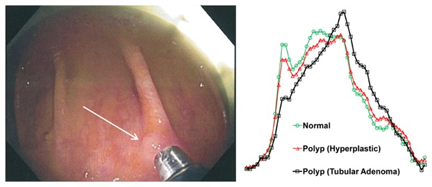
- Diffuse Reflectance Spectroscopy and Multi-spectral Imaging, which explores elastic light scattering and light absorption by tissue chromophores such as melanin, hemoglobin, bilirubin, and water.
- Fluorescence Spectroscopy and Imaging, which explore molecular electronic energy transition-related light emission of certain biomolecules in tissue such as tyrosine, tryptophan, NADH, collagen, and elastin.
- Raman Spectroscopy, which explores inelastic light scattering to give fingerprint-like spectral signatures of molecular vibrations. Molecules in tissue that have unique Raman signatures include proteins, DNA, lipids, glucose, and melanin.
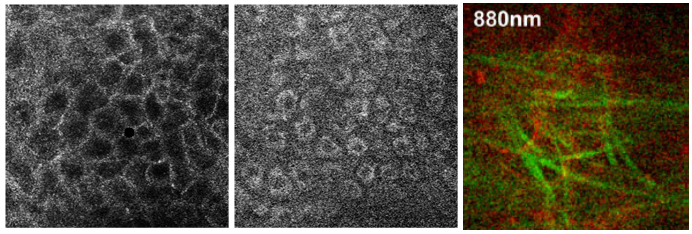
- In-Vivo Confocal Microscopy, which provides sectional images of tissue at micron spatial resolution, allowing us to visualize cellular structures and micro-circulation in real time on living tissue. Imaging mechanisms we are exploring include elastic scattering (reflectance), two-photon excitation fluorescence (TPEF), and second harmonic generation (SHG).
On the therapeutic side, we are focusing on developing applications for our new innovation – multiphoton photothermolysis. Based on the fact that multiphoton absorption occurs only at the focal point of a tightly focused femtosecond pulsed laser beam, this new therapeutic modality has precise spatial selectivity. Any targeting tissue microstructures seen under confocal/multiphoton microscopy can be precisely treated without affecting surrounding tissues. This “see and treat” theranostics approach has great potential for precision microsurgery treatment of diseases in complex organs such as the eye and brain, where high spatial selectivity is critical for preventing collateral effects on vision or central nervous system function.
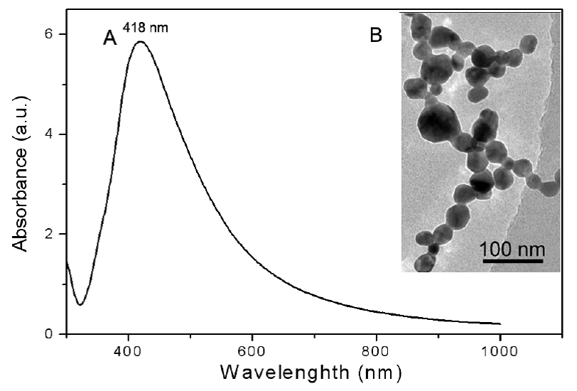
In addition, we are applying MEMS (micro-electro-mechanical systems)/ micromanufacturing technologies to miniaturize our optical devices for endoscopy uses and for point-of-care diagnostic applications. We are also applying nanotechnologies to enhance various light-tissue interactions for diagnostic and therapeutic applications, for example, using metal nanoparticle based SERS (surface enhanced Raman scattering) spectroscopy for diagnostics and developing nanoparticle based photothermal therapy.