The Molecular and Cellular Immunology Core (MCIC) is a full service facility specializing in advanced molecular and histological methods including multicolour immunohistochemistry (mcIHC) and immunofluorescence (mcIF), automated image analysis, laser capture microdissection, multispectral flow cytometry, and single-cell sequencing.
MCIC supports academic and industry projects both nationally and internationally, ranging from investigator-driven basic research, to correlative studies for clinical trials, to fee-for-service projects with industrial clients.
Molecular and Cellular Immunology Core
Multicolour immunohistochemistry
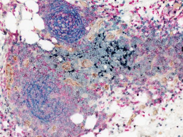
Tertiary lymphoid structure in high-grade serous carcinoma.
(CD21: blue, CD3: green, CD20: red, CD208: black, PNAd: brown,CD8: darker purple)
High dimensional flow cytometry
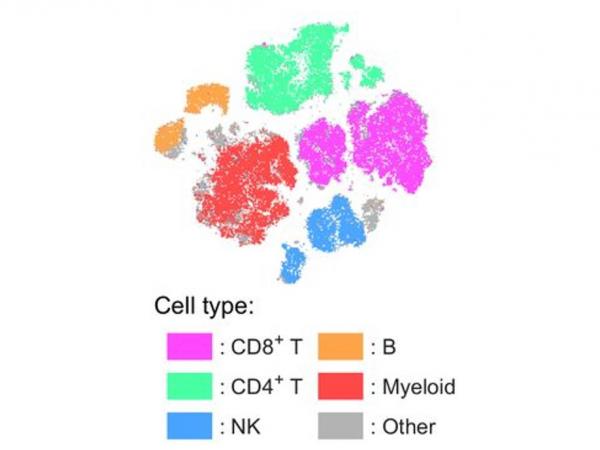
tSNE projections of immune cell compartments from disaggregated HGSC cells colored by cell type and defined using common phenotypic markers.
Laumont CM, Wouters MCA, Smazynski J, Gierc NS, Chavez EA, Chong LC, Thornton S, Milne K, Webb JR, Steidl C, Nelson BH. Single-cell Profiles and Prognostic Impact of Tumor-Infiltrating Lymphocytes Coexpressing CD39, CD103, and PD-1 in Ovarian Cancer. Clin Cancer Res. 2021 Jul 15;27(14):4089-4100. PMID: 33963000.
Multicolour immunofluorescence
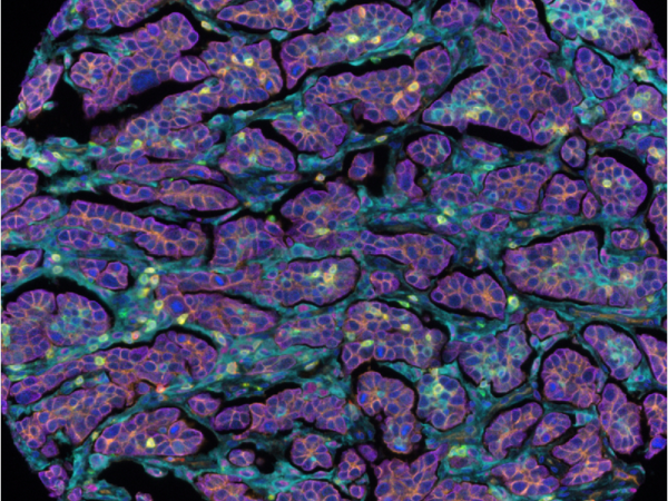
Tumour infiltrating lymphocytes in high-grade serous carcinoma.
(CD155: orange, CD68: magenta, PD-L1: cyan, CD3: yellow, CD8: green, PD-1: red, PanCK+: purple')
BC Cancer Foundation is the fundraising partner of BC Cancer, which includes BC Cancer Research. Together with our donors, we are changing cancer outcomes for British Columbians by funding innovative research and personalized treatment and care.